Breast Anatomy Quadrants / 1 / Read the blog to know what are the quadrants of the breast and preventive tips for breast cancer.. Lateral quadrants of the breast; Lymphatics from the left breast ultimately terminate in the thoracic duct and the left subclavian vein, and from the right breast in the right subclavian vein. Cancer of breast can spread to liver. The breast is somewhat circular in shape. The breast can be considered to be composed of two regions:
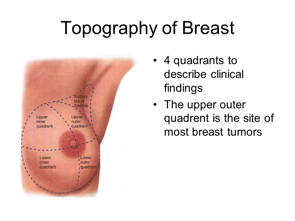
The outer quadrants of the breast are more likely than other sites to be injured, which can provoke damage to the glandular tissue and the development of a precancerous condition. Lymphatics from the left breast ultimately terminate in the thoracic duct and the left subclavian vein, and from the right breast in the right subclavian vein. Cancer of breast can spread to liver. Four quadrants of the breast • upper outer (superolateral) quadrant • upper inner (superomedial) quadrant • lower outer (inferolateral) quadrant. Applied lymph vessels of breast comunicates with those of abdomen.
Interventions For Clients With Breast Disorders Ppt Video Online Download from slideplayer.com Most breast cancers develop in the upper outer quadrant of the breast, closest to the armpit. The outer quadrants of the breast are more likely than other sites to be injured, which can provoke damage to the glandular tissue and the development of a precancerous condition. The breast can be considered to be composed of two regions: Cancer of breast can spread to liver. Breast tissue is drained by lymphatic vessels that lead to axillary nodes (which lie in the axilla) and internal mammary nodes (which lie along each side of the breast bone). Each lobe is comprised of many lobules, at the end of which are tiny bulb like glands, or sacs, where milk is produced in response to hormonal signals. Here is what we have learned from breast anatomy: There is a single tumor in two or more subsites and the subsite in which the tumor originated is unknown there is a single tumor located at the 12, 3, 6, or 9 o'clock position on the breast code the primary site to c509 when there are multiple tumors (two or more) in at least two quadrants of the breast
Learning anatomy is a massive undertaking, and we're here to help you pass with flying colours.
The breast tissue is usually distributed fairly symmetrically from left to right. Dominant pathway (receives >75% of lymph from breasts) drains lateral quadrants of breast either directly or via sappey's plexus to axillary nodes The major portion of the breast tissue is situated between the second and third rib superiorly, the sixth and seventh costal cartilage inferiorly, the anterior axillary line laterally, and the sternal border medially. Each breast contains 15 to 20 lobes arranged in a circular fashion. The upper outer quadrant extends towards the axilla as the axillary tail. Research has observed that the breast's outer upper quadrant is the most common site for the development of carcinoma of the breast, as seen in almost 60% of the cases. The breast is somewhat circular in shape. At the centre of the breast is the nipple, composed mostly of smooth muscle fibres. When breast cancer spreads, it is frequently to these nodes. Different quadrants of the breast A regional atlas of the human body, 5th ed., lippincott williams & wilkins, baltimore, md, 2006. Stromal tissue is the structural tissue of the breast includes fat and connective tissue. Each lobe is comprised of many lobules, at the end of which are tiny bulb like glands, or sacs, where milk is produced in response to hormonal signals.
It is a discussion about the internal structure of the breast and it finishes with a brief loo. The upper outer quadrant extends towards the axilla as the axillary tail. However, these ligaments are flexible and allow for movements in the breast. Here is what we have learned from breast anatomy: In 12 fresh female cadavers with no history of breast carcinoma, injections of patent blue dye were used to visualize the sns in the axillary quadrants and their lymphatic collectors from the upper outer quadrant of the breast, which is the most common location of breast cancer.
File Breast Quadrants Svg Wikimedia Commons from upload.wikimedia.org Lymphatics from the left breast ultimately terminate in the thoracic duct and the left subclavian vein, and from the right breast in the right subclavian vein. At the centre of the breast is the nipple, composed mostly of smooth muscle fibres. Variations in blood supply to the breast via these perforators explain why, in all quadrants of the breast, cancer has the potential to metastasize via parasternal lymphatics, especially in the deep medial aspect of the breast. Dominant pathway (receives >75% of lymph from breasts) drains lateral quadrants of breast either directly or via sappey's plexus to axillary nodes Quadrant wise drainage drainage from the 5 quadrants towards the axilla and internal mammary chain palpable + nonpalpable lesions axilla (%) imc (%) uoq 95.8 10.4 uiq 93.1 32.4 loq 97.7 29.5 liq 88.0 52.7 c 100 23.7 uoq: Breast tissue is drained by lymphatic vessels that lead to axillary nodes (which lie in the axilla) and internal mammary nodes (which lie along each side of the breast bone). The major portion of the breast tissue is situated between the second and third rib superiorly, the sixth and seventh costal cartilage inferiorly, the anterior axillary line laterally, and the sternal border medially. Applied lymph vessels of breast comunicates with those of abdomen.
Usually fibroglandular tissue occurs symmetrically in the upper outer quadrants of the breasts.
Applied lymph vessels of breast comunicates with those of abdomen. Lymphatics from the left breast ultimately terminate in the thoracic duct and the left subclavian vein, and from the right breast in the right subclavian vein. Learning anatomy is a massive undertaking, and we're here to help you pass with flying colours. Cancer registration & surveillance modules. Four quadrants of the breast • upper outer (superolateral) quadrant • upper inner (superomedial) quadrant • lower outer (inferolateral) quadrant. The breast is divided into four quadrants: Variations in blood supply to the breast via these perforators explain why, in all quadrants of the breast, cancer has the potential to metastasize via parasternal lymphatics, especially in the deep medial aspect of the breast. The upper outer quadrant extends towards the axilla as the axillary tail. Breast tissue is drained by lymphatic vessels that lead to axillary nodes (which lie in the axilla) and internal mammary nodes (which lie along each side of the breast bone). There is a single tumor in two or more subsites and the subsite in which the tumor originated is unknown there is a single tumor located at the 12, 3, 6, or 9 o'clock position on the breast code the primary site to c509 when there are multiple tumors (two or more) in at least two quadrants of the breast At the centre of the breast is the nipple, composed mostly of smooth muscle fibres. When breast cancer spreads, it is frequently to these nodes. Stromal tissue is the structural tissue of the breast includes fat and connective tissue.
Cancer registration & surveillance modules. Department of health and human services; Most breast cancers develop in the upper outer quadrant of the breast, closest to the armpit. The glandular parenchyma is estrogen dependent, thus on attaining menopause the glandular parenchyma atrophies. At the centre of the breast is the nipple, composed mostly of smooth muscle fibres.
Normal Breast Ultrasound How To from www.ultrasoundpaedia.com The glandular parenchyma is estrogen dependent, thus on attaining menopause the glandular parenchyma atrophies. The upper outer quadrant extends towards the axilla as the axillary tail. Research has observed that the breast's outer upper quadrant is the most common site for the development of carcinoma of the breast, as seen in almost 60% of the cases. There is a single tumor located at the 12, 3, 6, or 9 o'clock position on the breast code the primary site to c509 when there are multiple tumors (two or more) in at least two quadrants of the breast The pectoralis major muscle forms the base of the breast, which extends from the second to sixth rib early in life but may extend to below the sixth rib as the breast matures and sags. Four quadrants of the breast • upper outer (superolateral) quadrant • upper inner (superomedial) quadrant • lower outer (inferolateral) quadrant. It is a discussion about the internal structure of the breast and it finishes with a brief loo. Lateral quadrants of the breast;
Each of these 4 regions is called a quadrant.
Most breast cancers develop in the upper outer quadrant of the breast, closest to the armpit. Quadrant wise drainage drainage from the 5 quadrants towards the axilla and internal mammary chain palpable + nonpalpable lesions axilla (%) imc (%) uoq 95.8 10.4 uiq 93.1 32.4 loq 97.7 29.5 liq 88.0 52.7 c 100 23.7 uoq: Each of these 4 regions is called a quadrant. The breast is anchored to the pectoralis major fascia by the cooper ligaments. The pectoralis major muscle forms the base of the breast, which extends from the second to sixth rib early in life but may extend to below the sixth rib as the breast matures and sags. Different quadrants of the breast A layer of fatty tissue surrounds the breast glands and extends throughout the breast, which gives the breast a soft consistency and gentle, flowing contour. Each breast contains 15 to 20 lobes arranged in a circular fashion. The breast is a modified sweat gland located in the superficial fascia of the anterior chest wall. Mainly to the axillary lymph nodes as mentioned above. There is a single tumor in two or more subsites and the subsite in which the tumor originated is unknown there is a single tumor located at the 12, 3, 6, or 9 o'clock position on the breast code the primary site to c509 when there are multiple tumors (two or more) in at least two quadrants of the breast When breast cancer spreads, it is frequently to these nodes. The breast is somewhat circular in shape.


0 Komentar